
Using a complex, specially designed 3D printer, the scientists of regenerative medicine at Wake Forest Baptist Medical Center gave evidence that it is possible to print living tissues to replace injured or sick.
Scientists said that they created on the printer of ear, bone and muscle tissue. When implanted in animals, the forms have taken root and developed blood vessels. Most importantly, what do these early studies that the forms have the size you want and the function you wish to use them for humans.
Tissue engineering aims to regrow tissue and organs in the lab to help with the lack of donor tissue. Accurate printing in 3D enables the development of complex tissues and organs.
Tissue and Integrated Organ Printing System (ITOP), which was developed by engineers for ten years, supports both biological materials for the formation of tissues and water gels that hold cells. In addition, formed a strong temporary external structure. The printing process does not harm the cells.
One of the main goals of tissue engineering is the maintenance, implanted structures lived for a long time to integrate into the body. Scientists have addressed this in two ways. They have improved aqueous ink containing the cells so that those helped their health and growth, as well as printed circuit microchannels. These microchannels conduct nutrients and oxygen from the body in the printed form and keep them alive when they develop blood vessels.
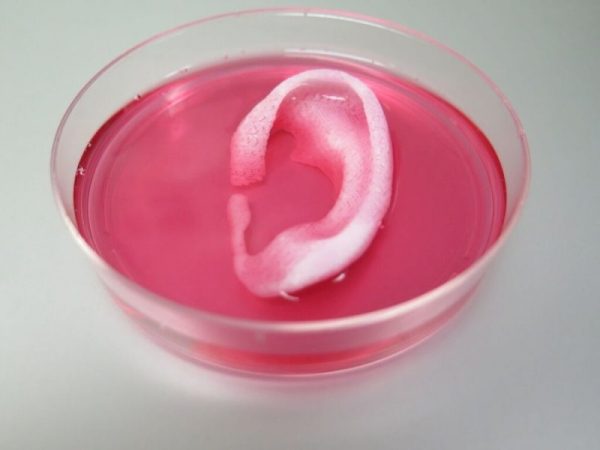
Ability ITOP demonstrated several experiments. To prove that ITOP can create complex 3D-printed form, the ears are of human size were embedded under the skin of rodents. Two months later the ear was in good condition and have developed cartilage and blood vessels.
To demonstrate that ITOP can create organized soft tissue structures, printed muscle tissue were implanted to rats. After two weeks of tests proved that the muscle remained strong and retained their abilities.
And to show the creation of the human bone structure size, the fragments of jaw bone were printed using human stem cells. The fragments had the size and shape required for reconstructing the faces of people. To study the maturation of bio-printed bone in the body, the printing segments are the bones of the skull were implanted in rats. After five months, the bio-printed patterns formed a well-developed vascular network in bone tissue.