 The work “pioneer” of medical photos.
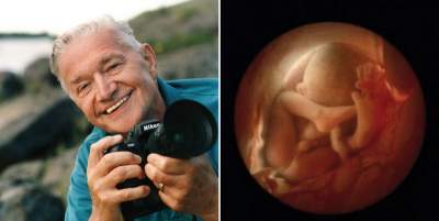
Since childhood, the microscope and the camera were the main Hobbies of a brilliant photographer Lennart Nilsson, who wanted to show the world the beauty of the human body at the cellular level. Pictures of the human fetus, Nilsson managed to get back in 1957, but they were not spectacular enough to show them to the public.
To obtain the most accurate and colourful pictures helped him a cystoscope is a medical instrument with which to examine the bladder from within. Nilsson attached the camera and light guide and made thousands of pictures of baby’s life inside the uterus.
So the skilled hands of Lennart Nilsson has created a miracle: revealed to the world the mystery of the origin of human life.
Lennart Nilsson was born 24 August 1922 Swedish city of Strangnas in a family that loved photography.
Even as a child Lennart was more interested in the microcosm, one that can only be seen with a microscope. Armed with a microscope and a camera, he could penetrate inaccessible to ordinary sight worlds, inner worlds of man in the truest sense of the word.
1.
The sperm in the fallopian tube

Its way into photography, Nielsen began in the mid-1940s, working as a freelancer for various Swedish publications. At this time, such works as “a Midwife in Lapland” and “Hunting for polar bears on Svalbard”, brought him international attention. The experiments in microphotography of Lennart starts in mid 1950-ies and actively cooperates with various scientific and medical organizations.
2.
Egg

First photograph of a human fetus, he succeeded in 1957. Unusual “candid” shot from the “bosom” of the female body became possible after Nelson after a series of experiments was able to combine the micro-camera and microspatial, pinning them to the tube of the cystoscope (the instrument used to inspect urinary bladder from the inside), so there is a unique shots that illustrate the process of formation of the human embryo and its development.
3.
Sperm

One of the 200 million paternal sperm, breaking through the shell of an egg, literally poured into her…
4.
Sperm

“When I first saw the fruit, he was 15 weeks and he was sucking the finger,” said Nielson. But the magazine editors wanted, so I removed the face of the fetus. It took a lot of years.”
International fame Nielson received in 1965 when LIFE magazine published 16 pages of photographs of a human embryo. These photos were immediately reproduced in Stern, Paris Match, The Sunday Times and other journals.
5.
The germ

In the same year he published a book of photographs Nilsson A Child is Born, the eight millionth edition which was sold out in the first few days. This book had several editions and is still one of the bestselling illustrated books in the history of this kind of albums.
6.
The fruit

In the future, Nielson continued their work, making not only photos but also movies.
In the 1960s — early 1970s, Nilsson collaborated with LIFE, making not only photomicrographs of different stages of fetal human development, but also other physiological processes in the organisms of humans and animals.
7.

The spacecraft Voyager I and Voyager II, carrying messages of extraterrestrial civilizations, in addition to other documents and photos and Nilsson. His scientific photographic work, he still continues.
8.
Week 8.

9.
10 weeks. The eyelids are already half open. Within a few days they will form completely.

10.

11.
16 weeks after fertilization. The skeleton consists mainly of flexible core and a network of blood vessels visible through the thin skin.

12.
16 weeks. The inquisitive toddler is already using their hands to explore the area.

13.
18 weeks. About 14 cm. the Fetus can now perceive sounds from the outside world.

14.
20 weeks after fertilization.

15.
26 weeks after fertilization

16.
36 weeks. In a month the baby will be born.

17.
Now the pioneer of medical photography Lennart Nilsson 91. Science and photography he has been so far. He’s a genius!

Related posts:
 Named the best photographs on EyeEm Photography Awards photo contest 2016. Photo
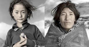
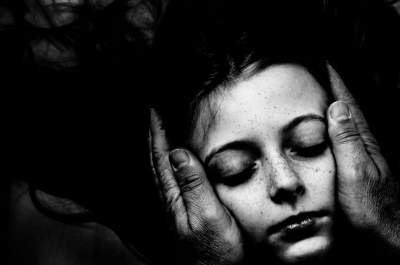
 The best pictures of the children at the competition, The B&W Child Photography 2016. Photo
 The best pictures of the children contest B&W Child Photography 2016. Photo
 The best works of the most famous women photographers. Photo